What is microdiscectomy for cervical fusion?

Anterior cervical fusion (ACDF) is a common and widely used surgical technique, which consists of replacing one or more cervical discs with a solid device, usually associated with a stabilizing element, with the intention of achieving immobilization of the operated segment and to decompress the medulla or nerve root compression.

What cases is it used for?

It is used for the treatment of both severe disc pathologies and cervical disc herniations causing cervicalgia (neck pain) and brachialgia (arm pain) or spinal compression (myelopathy). It can also be used for cervical instability as in spondylolisthesis, or severe cervical arthropathy causing chronic cervical pain.

What is involved in the operation?

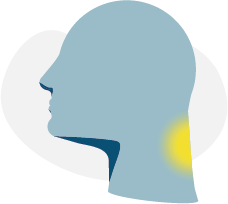
The surgeon makes a horizontal incision on one side of the neck to access the fascia of the platysma muscle. Immediately below is the sternocleidomastoid muscle (NDE), on the side, and the thyrohyoid, sternohyoid, and omohyoid muscles in medial position. These are carefully dissected, leading directly to the anterior vertebral space.

It is necessary to be very careful when working around the vasculo-nervous bundle (consisting of the carotid, jugular, and vagus nerve), the esophagus, and the medial airway. Finally, we find the prevertebral fascia, and flanking the vertebrae and discs, and the longus colli muscles, which we must separate to access the discs.

At this point, the microdiscectomy is carried out: using a microscope, the disc to be treated is cut and removed, until reaching the posterior common ligament, which is released, in order to visualize the dura that surrounds the medulla. This ensures that the compression on the medulla or nerve root has been removed.

With the help of an intraoperative fluoroscope, the appropriately sized interbody cage is placed, and in most cases a metal plate, anchored to the upper and lower adjacent vertebrae, then the sutures are performed.

In more complex cases in which the spinal compression is caused by the vertebra, the entire vertebral body can be removed and replaced with a larger device.

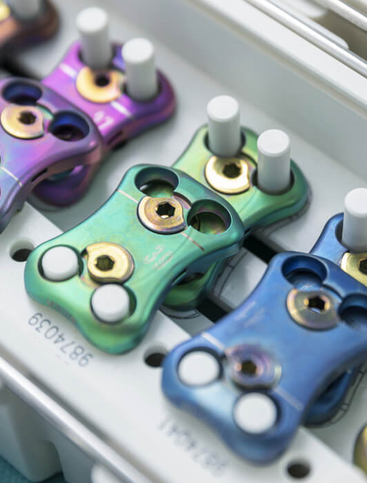
There are multiple types of interbody cages: solid or with bone graft, titanium or polymer (PEEK), with different types of anchorage. These must be adapted to the individual characteristics of each patient.

Recovery and rehabilitation

Since the total duration of the procedure is short and painless, the hospital stay ranges from 1 to 2 days. A soft cervical collar is required during the first few weeks, and specific rehabilitation should be performed in the following days to strengthen the muscles and regain range of motion. The rehabilitation time ranges from 4 to 8 weeks.

Risks of microdiscectomy for fusion

Complications during surgery are very rare. The main and most severe possible complication is hemorrhage, which can compress the airway and requires reoperation to be drained.

The most frequent postoperative complication is injury to the recurrent laryngeal nerve (the nerve which controls the muscles of the vocal cords), which is usually due to compression during surgery. It can affect the patient’s speech, causing aphonia, which in the vast majority of cases, resolves in the weeks following surgery. Microdiscectomy Spine Surgery in Delhi, India.

In the long term, cervical fusion of one or more levels can cause the adjacent discs to deteriorate more rapidly than they would just by natural aging. For this reason, in those cases in which there is no cervical instability or severe arthropathy, we may consider the option of cervical disc replacement with an artificial implant.

Would you like more information?
Tell us about your case!

Contact us so that we can give you a personalized assessment.

Share on:

©️ NuLyf Global Pvt. Ltd.